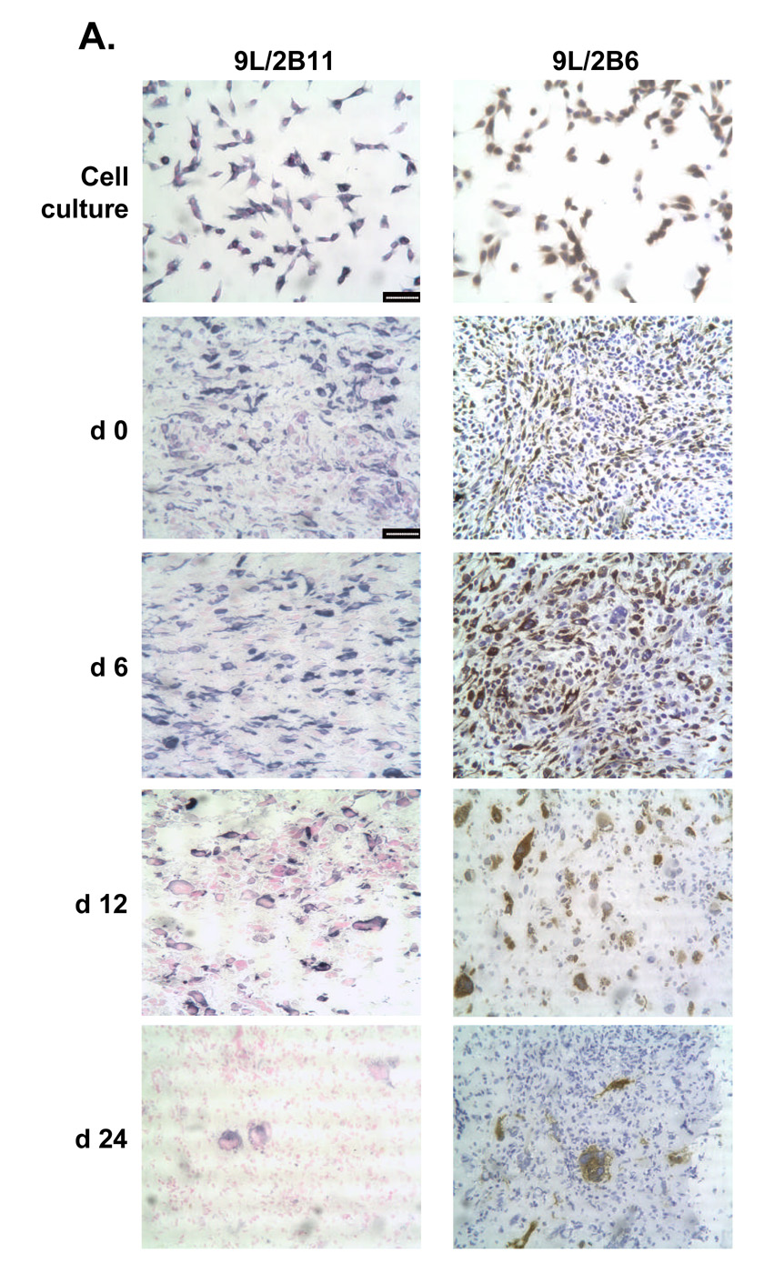
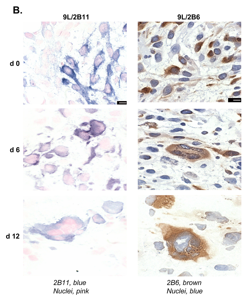
Figure 6. Reduced population of P450-positive cells and their morphological changes in 9L tumors.
A. Left panels, cultured 9L/2B11 cells (‘cell culture’) and 9L/2B11 tumor cryosections (day 0–24 after the first metronomic CPA injection) were immunostained with anti-P450 2B11 antibody (blue) and cell nuclei were labeled by Nuclear Fast Red (pink). Right panels, the corresponding set of cultured 9L/2B6 cells and 9L/2B6 cryosections were immunostained with anti-P450 2B6 antibody (brown) and cell nuclei were stained with hematoxylin (blue). The number of P450-positive cells decreased during metronomic CPA treatment, with re-population by P450-deficient cells evident on day 24, in particular in the case of the 9L/2B6 tumors. Scale bar, 50 µm. B. High magnification photos show enlarged P450 2B11 (left) and P450 2B6 (right) positive cells in sections of CPA-treated 9L/2B11 and 9L/2B6 tumors, respectively. These enlarged cells first appeared on day 6. Untreated tumor cells (day 0) are of normal size. Scale bar, 10 µm.


