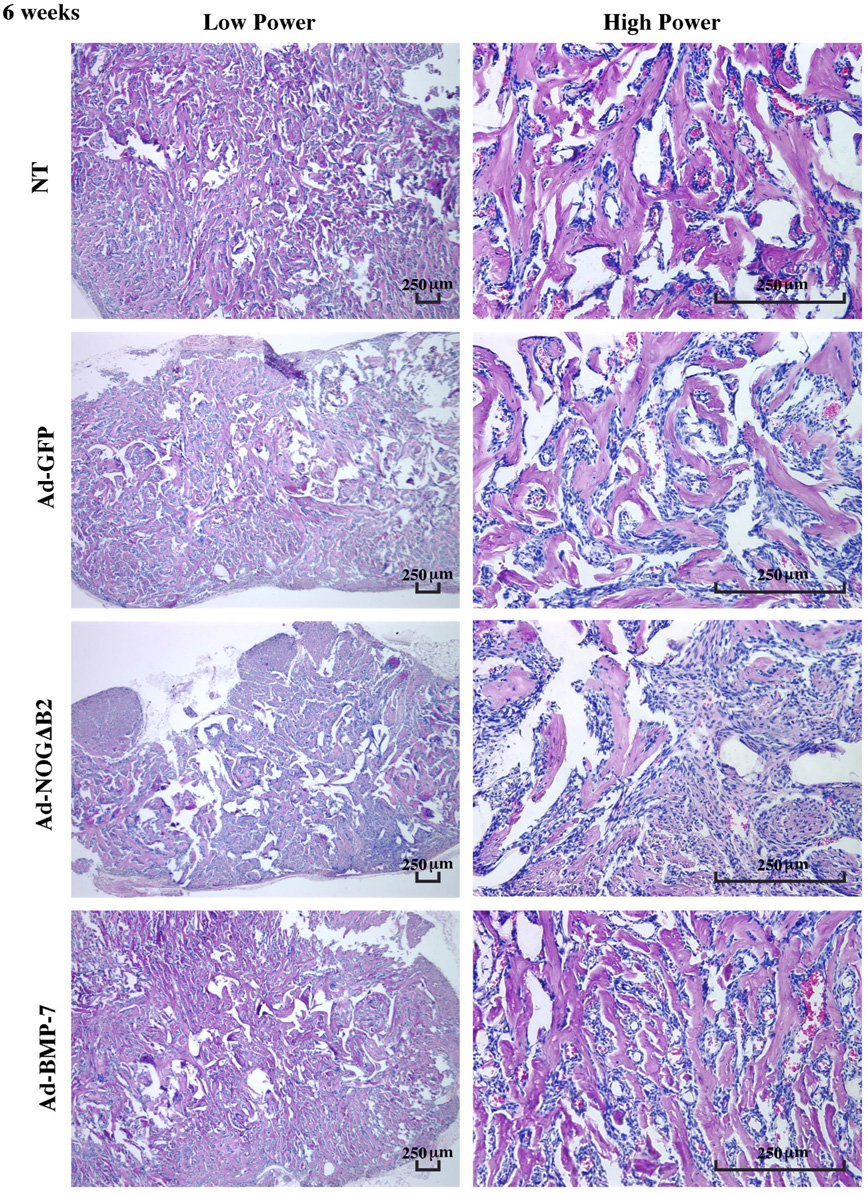
Figure 4.
Effect of BMP-7 and noggin gene transfer on mineralization in polymer scaffolds in vivo at 6 weeks. Left panels display low power photomicrographs; right panels show high power images. The mineral spicules became denser and more lamellar from 3–6 weeks. Noggin gene transfer decreased mineral area and implant size, while BMP gene transfer had minimal effects on mineralization compared with GFP treatment. Vascularization was formed among the mineral spicules. No bone marrow-like tissue was noted (H&E staining; bar = 250 µm; n = 3 animals/group).

