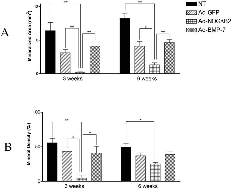
Figure 5.
Histomorphometric analysis of mineralized tissue area and density of the PLGA-cell implants at 3 and 6 weeks following gene transfer. Histomorpho-metric analysis was performed to measure. (A) Mineralized area of implants at 3 and 6 weeks. Implants from Ad-NOGΔB2 treatment of OCCM cells resulted in a reduction in mineral area compared with NT and Ad-GFP groups (*p < .05, **p < .01), while Ad-BMP-7 treatment had no effect on the induction of mineral. (B) Mineral density of implants from each group at 3 and 6 weeks. The mineral density of Ad-NOGΔB2 treated specimens were greatly reduced at 3 weeks compared with NT and Ad-GFP groups (*p< .05,** p <.01), at 6 weeks, Ad-NOGΔB2 treatment demonstrated slightly reduced mineral density when compared with NT (**p < .01), although it was not different compared with the implants from Ad-BMP-7 or Ad-GFP gene transfer.

