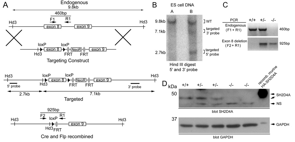
FIGURE 3.
Generation of sh2d4a gene-targeted mice. A, Shown is the organization of the endogenous sh2d4a locus, targeting vector and targeted sh2d4a allele before and after Cre and Flp-mediated recombination. Locations of HindIII restriction sites and 5’ and 3’ probes used in Southern blotting experiments and positions of PCR primers used in genotyping experiments are indicated. B, Shown is a Southern blot of HindIII digested genomic DNA from two ES cell clones electroporated with the sh2d4a targeting vector. The blot was probed with 5’ and 3’ probes simultaneously. Positions of bands from wild type and targeted sh2d4a alleles are shown. Clone B is correctly targeted. C, Mice that carried a germline targeted sh2d4a allele were crossed with actin-Flp transgenic mice to delete the NeoR cassette. Progeny were subsequently crossed with CMV-Cre transgenic mice to delete exon 8 of the sh2d4a gene in all tissues. Heterozygote NeoR-deleted sh2d4a exon 8-deleted mice were then intercrossed to generate wild type (+/+), heterozygote (+/−) and homozygote (−/−) NeoR-deleted sh2d4a exon 8-deleted progeny. Progeny were genotyped by PCR of tail DNA using the indicated primer combinations to detect wild-type and NeoR-deleted sh2d4a exon 8-deleted alleles. D, Expression of SH2D4A protein in whole splenocytes of littermate wild-type, heterozygote and homozygote NeoR-deleted sh2d4a exon 8-deleted mice was determined by Western blotting using an anti-SH2D4A antiserum. NS, non-specific. Note the absence of the 48 kDa SH2D4A band in the homozygous mutant animals. The membrane was re-probed with a GAPDH antibody to demonstrate equivalent protein loading.

