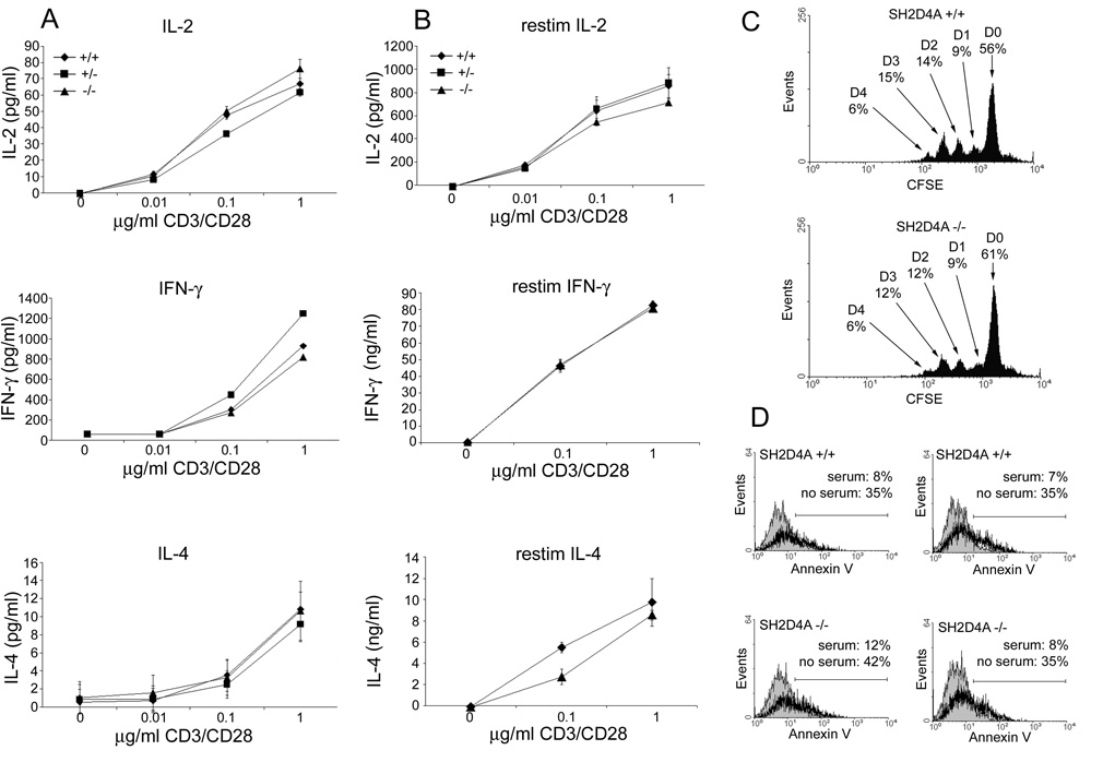
FIGURE 5.

T cell function in SH2D4A-deficient mice. A, Splenocytes from littermate wild-type, heterozygote or homozygote SH2D4A-deficient mice were stimulated with the indicated concentrations of a CD3 mAb and 0.5µg/ml of a CD28 mAb. Concentrations of IL-2 (at 24 h), IFN-γ and IL-4 (at 48 h) in culture supernatants were determined by ELISA. Data are represented as means +/− 1 SD of triplicate determinations. B, Splenocytes from littermate mice of the indicated genotypes were stimulated with CD3 and CD28 mAb for 48 h and then grown in IL-2 for a further 48 h. Cytokine synthesis in response to restimulation with CD3 and CD28 mAb was then determined as in A. C, Purified splenic T cells from littermate wild type and SH2D4A-deficient mice were stained with CFSE and stimulated with plate-bound CD3 mAb and 0.5µg/ml of soluble CD28 mAb. After 72 h, CFSE fluorescence was analyzed by flow cytometry. Indicated is the percentage of cells at successive cell division numbers, D0 through D4. D, Purified splenic T cells from littermate wild type and SH2D4A-deficient mice were stimulated with CD3 and CD28 mAb plus IL-2 for 48 h and then cultured in serum-containing (filled histogram) or serum-free (bold histogram) medium for a further 24 h. The percentage of Annexin-V-positive cells in cultures was then determined by flow cytometry. Shown are results from two different wild-type and SH2D4A-deficient mice.
