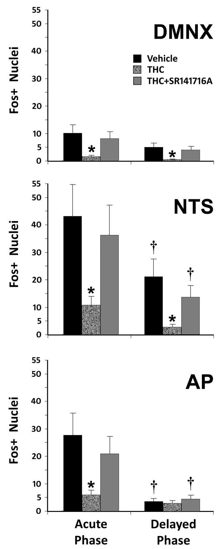
Figure 1.
Counts of Fos-immunoreactive Nuclei Following Acute or Delayed Phase Emesis in the Least Shrew. Fos-IR nuclei in the dorsal vagal complex (DVC) were analyzed for each nuclear region in the DVC. Induction of Fos (and vomiting) was through treatment with 10 mg/kg CIS (i.p.). Means and standard errors of Fos-IR nuclei are graphed for each pre-treatment condition, each subnucleus of the DVC, and both phases of CIV. Significantly reduced numbers of Fos-immunopositive nuclei (* p < 0.05) were noted after THC injection when compared to either the vehicle or SR141716A+THC conditions. When regions of the DVC having the same treatment conditions were compared between acute and delayed phases, the AP and NTS had significantly fewer († p < 0.05) Fos+ nuclei in the delayed phase than in the acute phase of emesis when treated with vehicle or SR141716A+THC, but not when treated with THC alone (p > 0.1). Numbers of Fos+ nuclei in the DMNX during the delayed phase were not significantly different from the acute phase (p > 0.12). Abbreviations: AP – area postrema; DMNX – dorsal motor nucleus of the vagus nerve; NTS – nucleus of the solitary tract.

