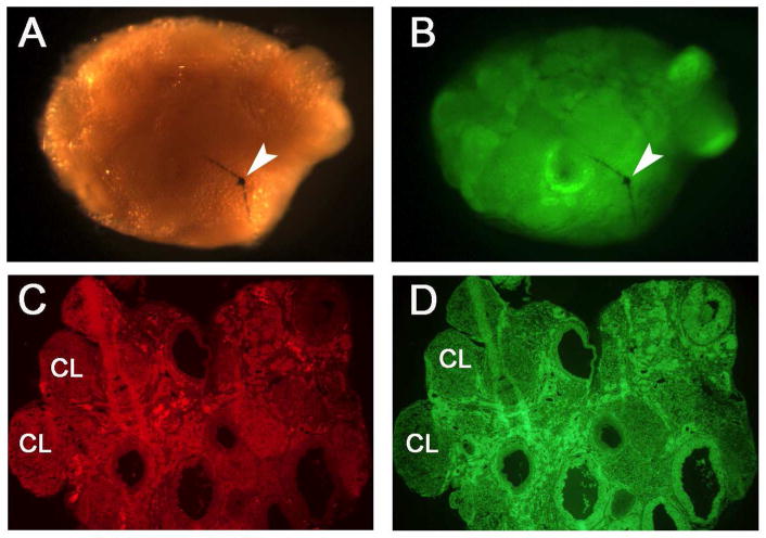
Fig. 2.
Examination of remnants (if any) of host ovary and morphology of transplanted ovaries 9 months post-transplantation in PND 8 groups. A representative ovary is shown using visible light (A) and UV light (B). To determine whether any remnant of host ovarian tissue remained, the ovaries were cleaned of surrounding bursa, oviduct, and fat tissues under a dissection microscope. The ovaries were then examined and imaged under visible light (A) and UV light (B) following the fixation as described in Materials and Methods. No or negligible host ovarian tissue remnants were observed in the recipient animals. Arrow indicates the suture used for closing the bursa ovary. Sections (8 μm) of quick frozen ovaries were prepared as described in Materials and Methods and used for determining the histology of the ovaries. The GFP ovaries were stained with EthD-2 and imaged using 550 nm (C; red) and 480 nm (D; green) filters. The ovary sections had different stages of the follicles including the corpus lutea (CL) at the time of collection. Original magnification of panel C and D is 40 x.

