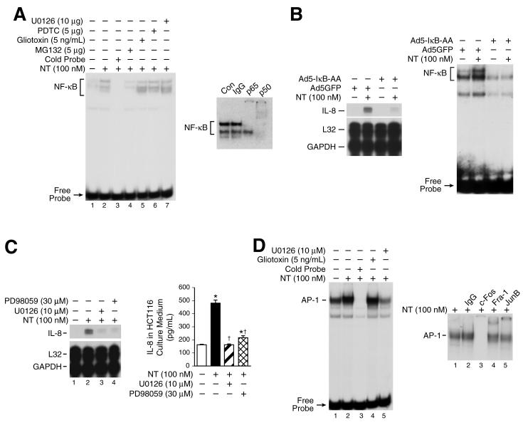
Figure 4. NF-κB activation and the MEK/AP-1 pathway play a role in IL-8 regulation by NT.
A. HCT116 cells were pretreated with vehicle or U0126 (10 μM), PDTC (5 μM), Giotoxin (5 ng/mL), MG132 (5 μM) for 25 min, and then treated with NT (100 nM) for 30 min. Cells were extracted for nuclear protein and analyzed by EMSA using a 32P-labeled NF-κB probe as described in “Materials and Methods” (left panel). Nuclear protein (10 μg per lane) from HCT116 treated with NT (100 nM) were preincubated with specific antibodies (p65 and p50) or IgG prior to the addition of 32P-labeled NF-κB probe and then DNA-binding activity was assessed by EMSA (right panel). B. HCT116 cells were infected with adenovirus encoding the super-repressor of IκBα (Ad5IκB-AA) or the adenovirus control vector encoding GFP (Ad5GFP) for 1h, washed and replated in McCoy’s 5A without FBS for 24 h and then treated with NT (100 nM) for 2 h. RNA was isolated and analyzed by RPA using the hCK-5 multi-probe (left panel). To confirm inhibition of NF-κB activation, cells were extracted for nuclear protein and analyzed by EMSA using a 32P-labeled NF-κB probe as described in “Materials and Methods” (right panel). C. HCT116 cells were pretreated with the MEK/ERK inhibitors PD98059 (30 μM) or U0126 (10 μM) for 20 min, and then treated with NT (100 nM) for 2 h; RNA was extracted and analyzed by RPA using the hCK-5 multi-probe(left panel). HCT116 cells were seeded in plates in McCoy’s 5A media with FBS. Twenty-four h later, media was changed to serum free and cells pretreated with PD98059 (30 μM) or U0126 (10 μM) at the indicated concentrations for 25 min, then treated with NT (100 nM) for 8 h. IL-8 secretion was measured in the conditioned media by ELISA(right panel). (Results are shown as mean ± SD and representative of three separate experiments; * = p < 0.05 vs control, † = p < 0.05 vs NT only). D. HCT116 cells were pretreated with U0126 (1 μM) or gliotoxin (5 ng/ml) for 25 min and then treated with NT (100 nM) for 30 min. Cells were extracted for nuclear protein and analyzed by EMSA using a 32P-labeled AP-1 probe as described in “Materials and Methods”(left panel). Nuclear extracts (10 μg per lane) from HCT116 cells were preincubated with specific antibodies (c-Fos, Fra-1 or JunB) prior to the addition of 32P-labeled AP-1 probe and then DNA-binding activity was assessed by EMSA(right panel).

