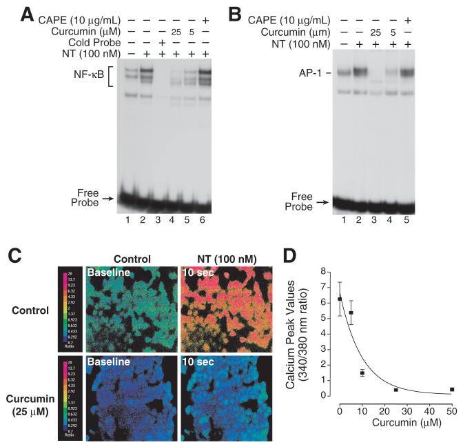
Figure 5. Curcumin inhibits NT-mediated IL-8 stimulation.
A. HCT116 cells were pretreated with CAPE (10 μg/ml) or curcumin (either 5 or 25 μM) for 25 min and then treated with NT (100 nM) for 30 min. Cells were extracted for nuclear protein and analyzed by EMSA using a 32P-labeled NF-κB probe as described in “Materials and Methods”. B. HCT116 cells were pretreated with curcumin (either 5 or 25 μM) or CAPE (10 μg/mL) for 25 min and then treated with NT (100 nM) for 30 min. Cells were extracted for nuclear protein and analyzed by EMSA using a 32P-labeled AP-1 probe as described in “Materials and Methods”. C. Pseudo-color images of HCT116 cells loaded with the Ca2+ sensitive dye, Fura-2. The cells were pretreated with vehicle or curcumin (25 mM) for 2 min, and then treated with NT (100 nM). The red color indicates the highest level of intracellular calcium while the blue-green color represents baseline levels. D. Pretreatment with increasing concentrations of curcumin decreases the peak [Ca2+]i response to NT (100 nM). (Each data point represents the average 340/380 nm ratio from 40 individual cells ± SD).

