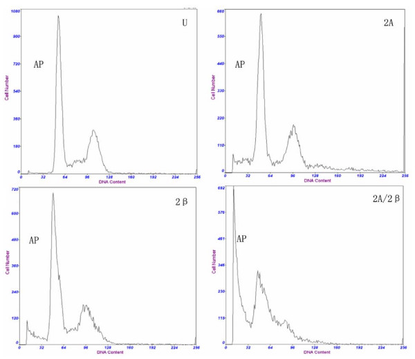
Figure 3.
shows that HepG2 cells without treatment (U), and were infected with LV-siMAT2A(2A), ALV-siMAT2β (2β)and LV-siMAT2A/2β (2A/2β), cell apoptosis was induced by LV-siMAT2A, ALV-siMAT2β and LV-siMAT2A/2β, as showed in the picture "AP" stands for apoptotic peak, There was most cells into apoptosis after cells were deal with LV-siMAT2A/2β, and G1/S arrest was discovered. It was demonstrated in the Fig. 3 that cell apoptosis caused by LV-siMAT2A/2β was increased in human hepatocelluar cancer cells HepG2 compared to that induced by LV-siMAT2A, ALV-siMAT2β (P < 0.01) and control, but they all had no difference between LV-siMAT2A and LV-siMAT2β.

