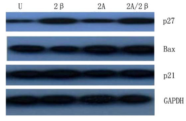
Figure 5.
showed that HepG2 cells without treatment (U), and were infected withLV-siMAT2β (2β), A LV-siMAT2A(2A)and LV-siMAT2A/2β (2A/2β), LV-siMAT2β, ALV-siMAT2A and LV-siMAT2A/2β. Western-blot was applied to assay the expressions of Bax, p27 and p21 GAPDH was used as control, It was showed in Fig 8 that protein of Bax was increased by LV-siMAT2β, ALV-siMAT2A and LV-siMAT2A/2β, which was highest after treating with LV-siMAT2A/2β, protein of p27 was increased by LV-siMAT2β and LV-siMAT2A/2β but not by LV-siMAT2A, The expression of p21 does not change.

