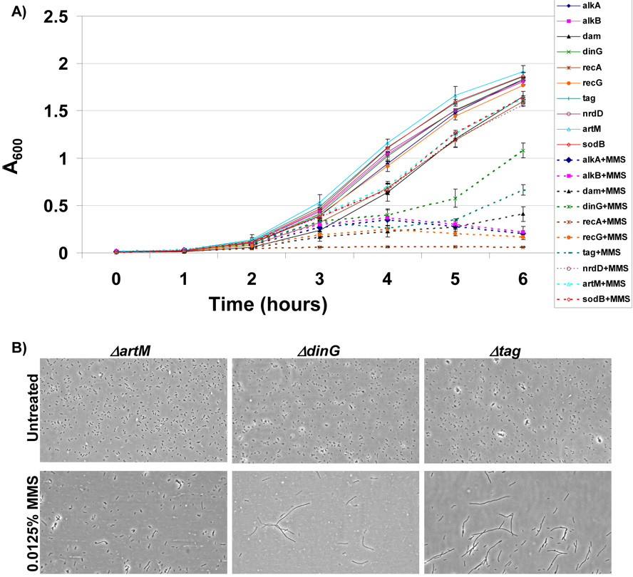
Figure 5. Measures of Growth and Filamentation in Identified MMS-Sensitive Mutants.
(A) The growth of MMS sensitive mutants identified by microarray analysis and wild-type surrogates was analyzed in LB-chloramphenicol −/+ 0.015% MMS. The growth of individual gene deletion mutants in untreated (solid lines) and MMS-treated media (dashed lines) was measured spectrophotometrically (A600) and plotted as a function of time. The six hour time point coincides with the endpoint used for microarray analysis and demonstrates that each of the MMS sensitive mutants identified by microarray analysis (ΔalkA, ΔalkB, Δdam, ΔdinG, ΔrecA, ΔrecG, and Δtag) was growth impaired in MMS containing media. (B) The cellular morphology of the MMS sensitive gene deletion mutants (ΔdinG and Δtag) and a wild-type like control (ΔartM) were analyzed from samples grown for 5 hours in LB-chloramphenicol −/+ 0.015% MMS. Cells were imaged on a Nikon TS-100 microscope at 40X magnification and demonstrated that ΔdinG and Δtag cells filament in response to MMS damage.

