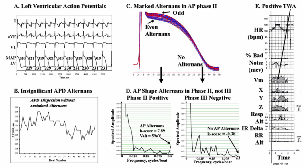
Figure 2.
Alternans Predominates in AP Phase II of Left Ventricular APs in this 59 year old man with coronary disease and LVEF 21%. (A, B) APD does not oscillate during pacing. However, (C ) Marked AP amplitude alternans in phase II is seen (red/blue separation). (D) AP alternans occurs in phase II (arrowed: k-score=7.9, Valt=59µV) but is undetectable in phase III (k-score −0.38); APD variations were also minimal. (E) TWA was positive at this time (arrowed) in leads Y, Z and Vm. Same abbreviations as figure 1.

