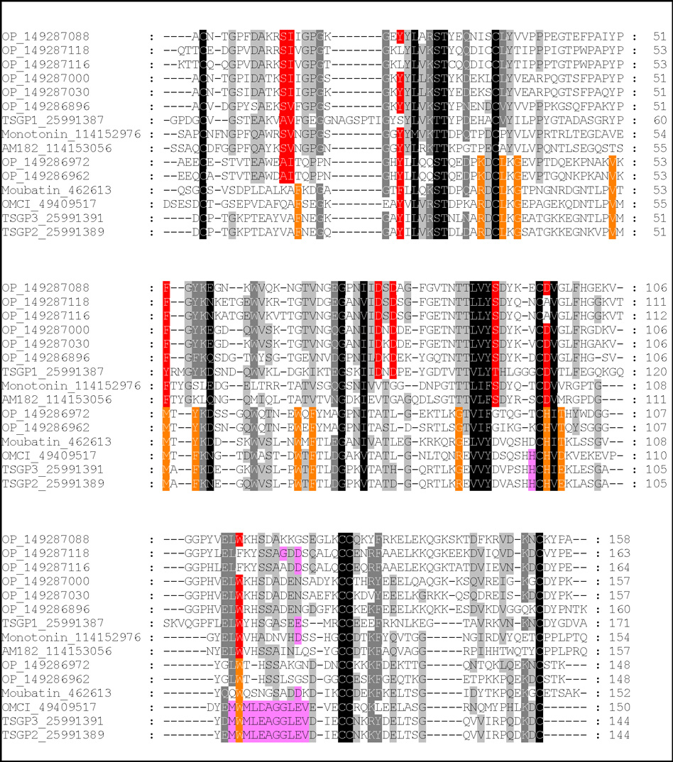
Fig. 10.
Multiple alignment of tick lipocalins from the moubatin and serotonin and histamine-binding clades. Conserved residues that line the ligand binding pocket of the moubatin-clade lipocalins are colored in green. Binding pocket data were inferred from those residues that interact with ricinuleic acid in the structure of OMCI (Roversi et al. 2007). Conserved residues that interact with serotonin and histamine are colored in yellow. Data were obtained from the structures of monotonin (Mans et al. 2008). Residues implicated in complement C5 interaction are colored purple.

