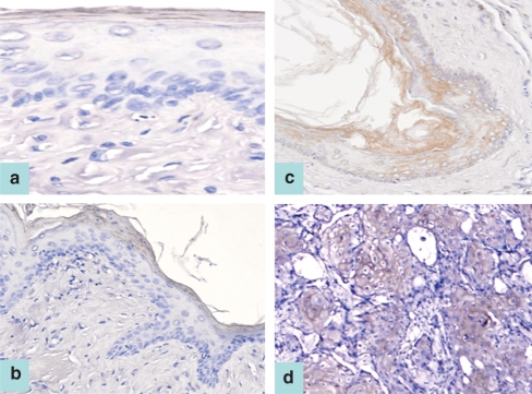
Figure 2.
cIAP1 staining was located in the outermost keratinized layer of an untreated pouch tissue specimen (a, ABC stain ×200), as well as a representative 3-week 7,12-dimethylbenz[a]anthracene (DMBA)-treated pouch tissue (b, ABC stain ×100). Patchy cytoplasmic staining was detected in the whole layer of a representative specimen of 7-week DMBA-treated pouch tissue (c, ABC stain ×100). A representative specimen of pouch tissue treated with DMBA for 14 weeks demonstrated cytoplasmic cIAP1 staining (d) (ABC stain ×100). A similar immunostaining pattern is also observed for XIAP and NAIP proteins.

