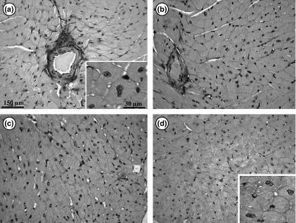
Figure 7.
Photomicrographs showing the left ventricle myocardium in transgenic mice. (a) Ex-M: exercised mice treated with mesterolone; (b) Ex-C: exercised animals treated with gum arabic (vehicle); (c) Sed-M: sedentary animals treated with mesterolone; (d) Sed-C sedentary mice treated with vehicle. Note that qualitatively cardiomyocytes are higher in size in exercised (a, b) and smaller in sedentary (d, c) groups. The insets depict a high magnification of cardiomyocytes of Ex-M (a) and Se-C (d).

