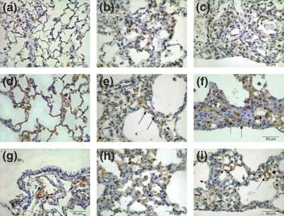
Figure 2.
Control group shows the uniform distribution and minimal proportion of SP-A positive cells in alveolar septa (a). In the early BHT (b) group, a homogeneous distribution along the alveolar wall contrasts with a non-homogeneous SP-A profile, poorly distributed along the collapsed and fibroblastic foci areas found in late BHT (c) lungs. Microvasculature immunostaining for factor VIII in the control (d) group shows the uniform distribution coincident with the maintenance of pulmonary architecture. The early BHT (e) group shows diffuse and poor microvasculature density, which is highly active and coincident with the inflammatory alveolar septal thickening. In the late (f) BHT group, a heterogeneous and poor microvasculature density, which is highly active, coincides with the distortion of the pulmonary architecture. VCAM-1 endothelial adhesion molecules in the control (g) is poor, in contrast with significant increase in early (h) and late (i) BHT lungs. Immunostaining for SP-A (a,b,c), factor VIII (d,e,f) and VCAM-1 (g,h,i) was carried out at 400×.

