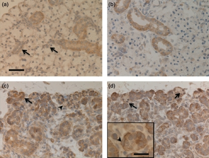
Figure 1.
Bcl-2 immunohistochemistry. Bar = 50 μm (a) Control. Striated duct cells are positive. Intercalated duct cells (arrows) and acinar cells are negative. (b) Day 1: Cells in dilated ducts show positive reaction. (c) Day 5: The remaining duct cells are immunoreactive to Bcl-2 antibody. There are some positive (arrow) and many negative (arrowhead) acinar cells in the atrophic gland. (d) Day 14: Most residual duct cells are positive and remaining acinar cells are negative (arrows). Inset: higher magnification of (d). Bar = 20 μm. The spindle-shaped (myoepithelial-like) cell is immunopositive (arrowhead).

