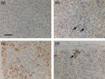
Figure 2.
Bax immunohistochemistry. Bar = 50 μm (a) Control. No immunoreaction is identified in normal submandibular glands. (b) Day 1: There are some positive acinar cells (arrows). (c) Day 3: Many acinar cells are Bax immunoreactive. (d) Day 14: The remaining acinar cells are reactive with the anti-Bax antibody (arrow).

