
Figure 1.
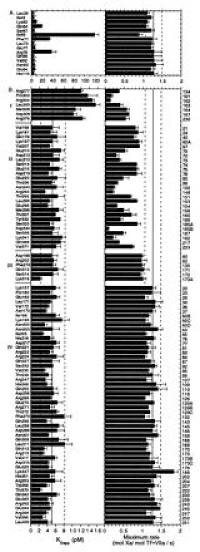
Affinity for phospholipid reconstituted TF (5 pM) (Left) and the maximum rate of Xa formation (Right) are shown for individual Ala replacement mutants at residue positions in the light chain (A) and the protease domain (B) of VII. Solid and dashed lines indicate mean ± 2 SD, respectively, for values obtained with wild-type VII transiently expressed in parallel with mutant VII. Protease domain mutants are grouped according to the functional defects: (I) combined decrease in TF binding and proteolytic function, (II) selective loss of proteolytic function to >3 SD of control, (III) subtle loss of proteolytic function (<3 and >2 SD), and (IV) normal function. Mean ± SD (n ≥ 3) are shown. In the right margin, chymotrypsin numbering is given for the protease domain. Replacement of the following residues resulted in maximum rates <0.1 s−1, and these essentially inactive mutants are not included in the figure: Leu-177 [c41], Leu-287 [c144], Ser-344 [c195], Val-362 [c213], and Trp-364 [c215].