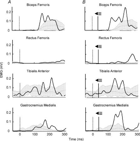
Figure 2. Examples of EMG responses for obstacle avoidance.
EMG activity of biceps femoris, rectus femoris, tibialis anterior and gastrocnemius medialis in response to an obstacle release at mid-swing. Representative trials from one subject. A, no startle trial. B, startle trial. The vertical dotted line indicates the obstacle release moment. The vertical continuous line shows when the startle was given. The shaded area represents mean and ±.2 s.d.s of EMG activity of the control stride. Superimposed (continuous line) is the trace of the representative trial. The obstacle was released at 72.8% of the step cycle in A and at 71.1% of the step cycle in B, which accounts for the slight delay of the control stride in B with respect to A (difference of 20 ms).

