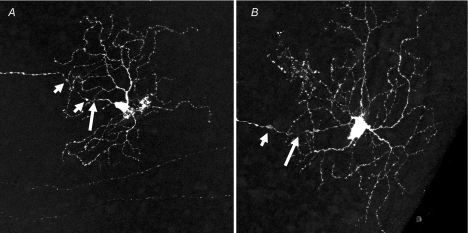
Figure 1. Retinal ganglion cell abnormalities in retinas from diabetic Ins2Akita mice.
Ins2Akita and Thy1-YFP mice were crossbred to produce spontaneously diabetic animals with endogenous expression of the yellow fluorescent protein in a subset of RGCs. Cells were imaged by confocal microcopy (Leica TCS SP2 AOBS) and rendered as maximum projections from z-stacks that included the axon and entire dendritic field. Abnormal features were noted in the ganglion cells of retinas from mice that were diabetic for three months. The abnormalities included axonal swellings (short arrows) and associated constriction (long arrows), as well as enlarged cell bodies and increased dendritic branches and terminals. A, medium ON-RGC; B, large ON-RGC (Gastinger et al. 2008).

