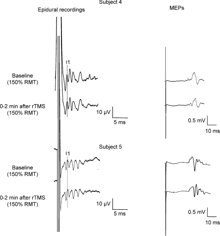
Figure 2. Corticospinal volleys and motor-evoked potentials evoked by single pulse magnetic stimulation in baseline conditions and after 1 Hz rTMS (900 stimuli) in subjects 4 and 5.
Each trace is the average of 20 sweeps. Magnetic stimulation evokes four descending waves in patient 4. The peak of the earliest (I1) I-wave is indicated by the vertical line. Two minutes after rTMS, the size of the later I-waves is decreased, the amplitude of the I1-wave is unchanged. The suppression is more pronounced for the last I-wave (I4). The amplitude of MEP is also decreased after rTMS. Magnetic stimulation evokes five descending waves in subject 5. The peak of the earliest (I1) I-wave is indicated by the vertical line. Two minutes after rTMS, the size of the later I-waves and of MEPs is unchanged.

