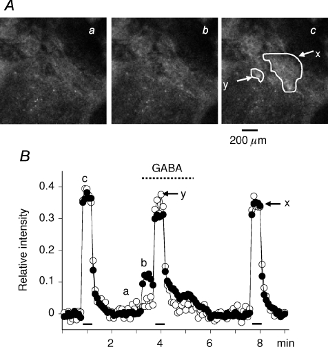
Figure 9. Inhibitory effects of GABA on trans-synaptically evoked Ca2+ response in rat AM cells.
A, confocal images of fluo-4 fluorescence in rat adrenal medulla. The adrenal gland was retrogradely perfused through the adrenal vein with saline (see Methods). GABA at 30 μm was added to the perfusion solution during the indicated period (interrupted line). Nerve fibres remaining in the gland were electrically stimulated with 60 V pulses of 1.5 ms duration at 10 Hz for 30 s during the indicated periods (bars). The adrenal medulla was illuminated with 488 laser and emission of above 510 nm was observed every 5 s. B, relative values of change in fluorescence intensity in the presence and absence of GABA are plotted against time. Fluorescence intensities in the areas (x and y) indicated in Ac were measured and presented as filled (x) and open (y) symbols, respectively. After correction for the decline due to photobleaching, an increase in fluorescence intensity in response to electrical stimulation and GABA was expressed as a fraction of the resting level (see Methods). a, b and c in A correspond to a, b and c in B.

