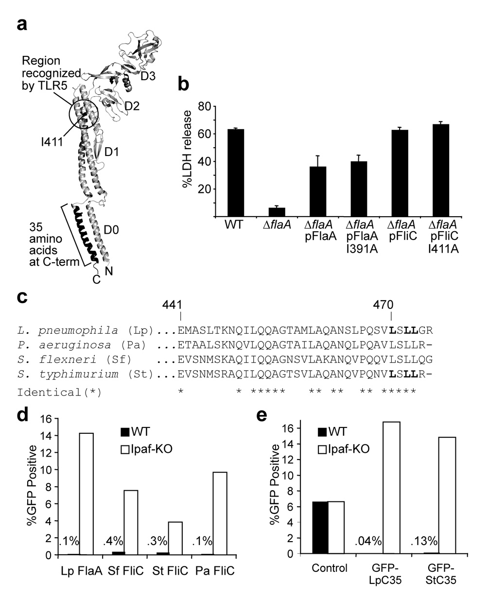
Figure 2. The cytotoxic C-terminus of flagellin is highly conserved and is distinct from the region sensed by TLR5.
(a) Model of the salmonella flagellin monomer as it appears within the assembled flagellar filament40. Isoleucine 411 is critical for ‘sensing’ of flagellin by TLR539, and corresponds to I391 in L. pneumophila flagellin. (b) Cell death assayed by release of lactate dehydrogenase (LDH) from B6 macrophages infected with indicated L. pneumophila strains (MOI = 1). Error bars represent s.d. of triplicate samples. (c) Alignment of the C-terminal 35 amino acids of flagellin; bolded leucines in Lp and St flagellin were mutated to alanine in the experiments in Fig. 3. (d,e) Graphs of flow cytometry data on wild-type (WT) or IPAF-deficient (IPAF-KO) macrophages transduced with retroviruses expressing either full-length flagellin from L. pneumophila (Lp-FlaA), S. typhimurium (St-FliC), P. aeruginosa (Pa-FliC) or S. flexneri (Sf-FliC) upstream of IRES-GFP (d) or the C-terminal 35 amino acids (C35) of L. pneumophila (Lp) or S. typhimurium (St) flagellin fused to the C-terminus of GFP (e). The percentage of macrophages that were GFP+ is indicated. Results are representative of 2–5 independent experiments.

