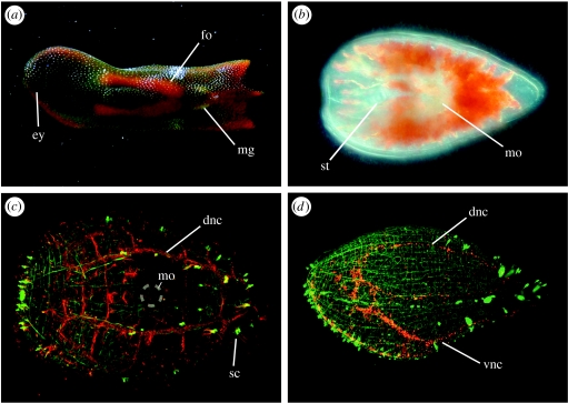
Figure 3.
Morphology of the acoel C. longifissura. (a) Adult with ripe female (fo) and male genital (mg) organs. A pair of eyes (ey) is located at the anterior end. (b) At hatching, the juvenile possesses a statocyst (st) and a pair of lateral eyes. The mouth opening (mo) is ventral, anterior to the left. (c) Confocal image of a juvenile to visualize the nervous system. Actin is visualized with Alexa-488 phalloidin (green) and microtubules with anti-tubulin antibody (red). Dorsal view, the nervous system runs orthogonally with bilateral nerve chords on the dorsal and ventrolateral side (dnc, vnc), sensory cells (sc) are connected with the main nerve chords. The muscular system is composed out of longitudinal and circular musculature. The position of the mouth opening (mo) is indicated with a circle. (d) Lateral view of a juvenile (green phalloidin, red anti-serotonin). The serotonergic subset of the nervous system is labelled in red.

