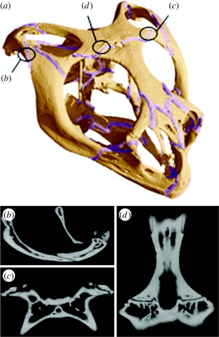
Figure 1.
Cranial sutures of Uromastyx. (a) All the sutures represented in the high-resolution full suture model and the locations of the individual model sutures, (b) a lateral view of a micro-computed tomography image showing the jugal–squamosal suture, (c) a ventral view of the postorbital–parietal suture and (d) a ventral view of the frontal–parietal suture.

