
Figure 3. Immunocytochemical analysis for nuclear Smad2 and Smad4 protein.
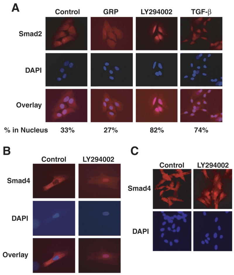
(A) GRP treatment resulted in a slightly decreased level of nuclear Smad2 protein. Inhibition of PI3-K by LY294002 significantly increased nuclear accumulation of Smad2, which was greater than treatment with TGF-β (1 ng/ml) alone. Quantitative Smad2 nuclear localization is expressed as percentage of Smad2-stained cells relative to total number of cells examined. (B and C) Similarly, LY294002 treatment resulted in intense nuclear staining of Smad4.