Figure 3.
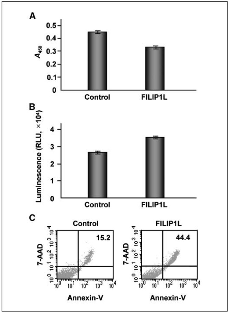
Overexpression of FILIP1L in endothelial cells leads to inhibition of cell proliferation and an increase in apoptosis. A, inhibition of cell proliferation by overexpression of FILIP1L in HUVECs was analyzed by BrdUrd ELISA 24 h after transfection. The amount of BrdUrd incorporated was measured by absorbance at 450 nm. Bars, SE (n = 4, P < 0.0001). The result is representative of three independent experiments. B, increased apoptosis by overexpression of FILIP1L in HUVECs was analyzed by caspase-3/caspase-7 assay 24 h after transfection. Caspase-3/caspase-7 activity was measured by luminescence. Bars, SE (n = 4, P < 0.001). The result is representative of three independent experiments. C, increased apoptosis by overexpression of FILIP1L in HUVECs was analyzed by Annexin V–FITC and 7-AAD staining followed by flow cytometry analysis 48 h after transfection. The numbers 15.2 for control and 44.4 for FILIP1L indicate the percentage of cells in late apoptosis. The result is representative of two independent experiments.

