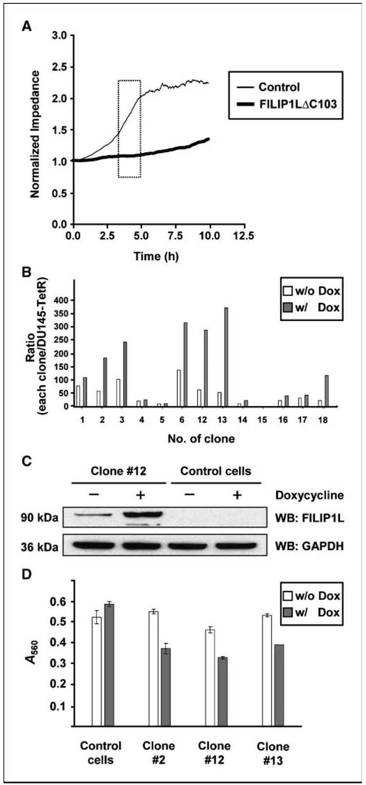
Figure 5.
Overexpression of FILIP1LΔC103 in HUVECs, as well as DU145 prostate cancer cells, leads to inhibition of cell migration. A, FILIP1LΔC103-transfected HUVECs showed a significantly slower migration rate than control vector-transfected HUVECs as measured by Electric Cell-Substrate Impedance Sensing System in real time (P < 0.0001). Square box, linear range in the curve that was used for analysis. The result is representative of three independent experiments. B, real-time RT-PCR analysis for FILIP1L on cDNA from DU145 clones transduced with FILIP1LΔC103-lentivirus. Each clone was treated with either PBS (w/o Dox) or 1 µg/mL doxycycline (Dox) for 48 h before harvest. The y axis represents a ratio between each clone and the parental Tet repressor-expressing DU145 cells where each value was standardized with housekeeping gene GAPDH. Columns, average of three experiments. C, a 90-kDa FILIP1LΔC103 protein was detected in Tet repressor-expressing DU145 cells transduced with FILIP1LΔC103-lentivirus, but not control lentivirus, by Western blot using anti-FILIP1L antibody. The result shown is a representative (clone 12) from several clones. GAPDH blot is shown as the loading control. D, all three FILIP1LΔC103 clones, but not mixed population of control cells, showed a significantly slower migration in the presence of doxycycline. Error bars indicate SE (n = 3). P value comparisons between in the presence and in the absence of doxycycline were as follows: control cells, P = 0.141; clone 2, P = 0.0014; clone 12, P = 0.0005; and clone 13, P < 0.0001. The result is representative of two independent experiments.

