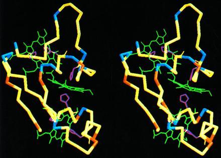
Figure 5.

Stereoview of backbone and hemes of the energy-minimized average structure of D. acetoxidans Cyt c7. The positively and negatively charged residues are shown in blue and red, respectively. The neutral residues are shown in yellow. The three-heme groups are green, and the axial histidines are yellow. The molecule orientation is the same as in Fig. 3.
