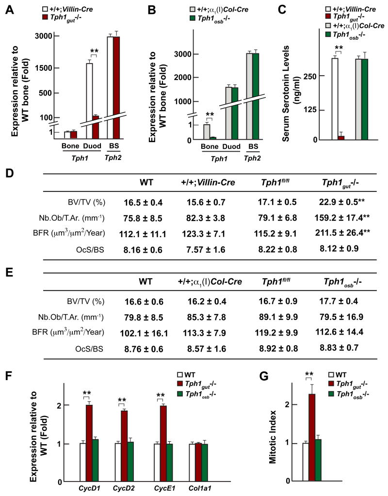
Figure 4. Duodenal-derived serotonin regulates bone formation.
(A–B) Real-time PCR analysis of Tph1 expression in gut and long bones (Bone) and Tph2 expression in the brainstem (BS) of Tph1gut−/− and Tph1osb−/−compared to +/+;Villin-Cre and +/+;α1(I)Col-Cre mice respectively.
(C–E) Serum serotonin levels (C) and bone histomorphometric analysis (vertebrae) (D–E) in WT, +/+;Villin-Cre, Tph1gut−/−, +/+;α1(I)Col-Cre and Tph1osb−/− mice.
(F) Real-time PCR analysis of Cyclins and Col1a1 expression in long bones of WT, Tph1gut−/− and Tph1osb−/− mice.
(G) In vivo osteoblast proliferation in WT, Tph1gut−/− and Tph1osb−/− mice.

