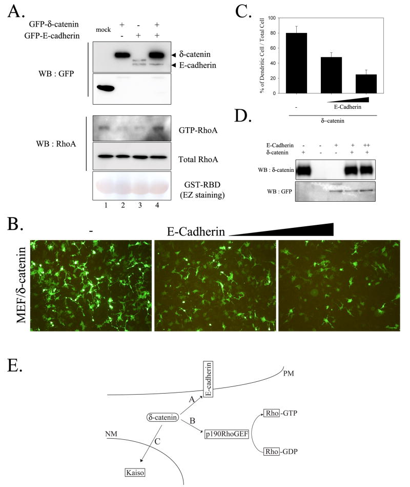
Figure 4. Co-expression of E-cadherin with d-catenin reduced the effects of d-catenin on lowering RhoA activity and dendrite-like process formation.
(A) Levels of GTP-RhoA were measured in MEF cells transfected as indicated. (B) MEF cells were transfected with d-catenin or increased amount of E-cadherin expressing plasmid, and fluorescent image was taken after 1-day post-transfection. (C) Percentage of cells showing dendrite-like process formation among total GFP-positive cells. (D) MEF cell lysates were subjected to western blotting and levels of d-catenin and E-cadherin were shown. (E) Schematic illustration of δ-catenin interaction with different binding partners in cells. A) In the presence of E-cadherin and/or E-cadherin-mediated cell-cell contacts, cellular microenvironment is preferential for δ-catenin localization in the plasma membrane and for the association of δ-catenin with E-cadherin. B) In the absence of E-cadherin and/or E-cadherin-mediated cell-cell contact, δ-catenin is localized in the cytoplasm and binds cytoplasmic proteins such as p190RhoGEF, which results in the down-regulation of RhoA activity. C) δ-catenin enters the nucleus and interacts with Kaiso, a transcription factor. The signal for δ-catenin nuclear localization and/or function remains elusive. PM, plasma membrane; NM, nuclear membrane.

