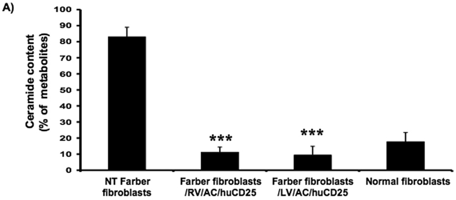
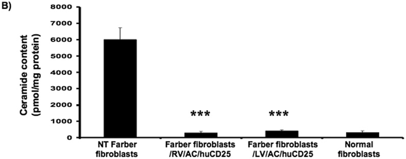
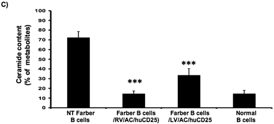
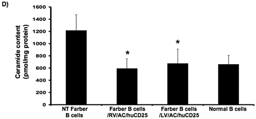
FIG. 2. Lysosomal ceramide turnover and ceramide content in transduced Farber patient cells.
Immortalized Farber patient cells were transduced with either oncoretrovirus (RV) or lentivirus (LV) encoding human AC and huCD25. Non-transduced and transduced Farber patient cells were pulsed with [3H-ceramide]-sphingomyelin for 48 h. Lipids were isolated, and then separated by TLC. The AC activity of fibroblasts (A) and B cells (C) are shown. To determine ceramide content, lipid extracts were incubated with E. coli diacylglycerol kinase and [γ32P]ATP. Radioactive ceramide 1-phosphate was isolated by TLC and quantified by liquid scintillation analysis for both fibroblast (B) and B cell (D) extracts. Error bars represent SD; measurements are averages of at least three separate experiments. * p < 0.05, *** p < 0.001, for groups indicated versus non-transduced (NT) controls.


