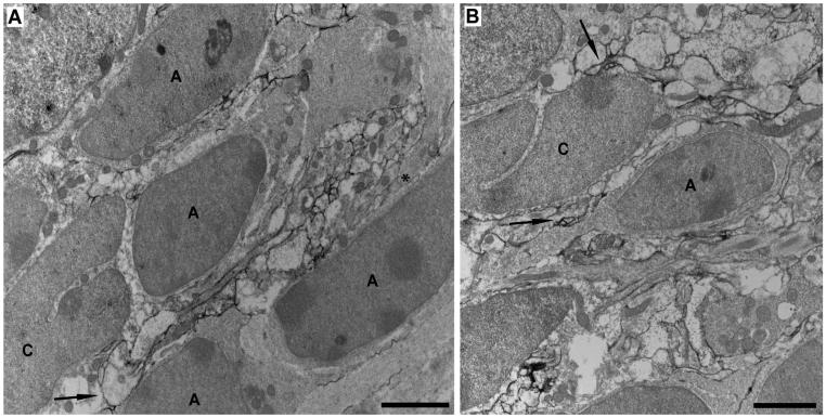
Figure 5.
A,B: Preembedding NG2 immunoelectron microscopy of the SVZ in a P30 mouse using a rabbit anti-NG2 antibody against the ectodomain of the NG2 core protein. NG2-immunolabeled section without lead or uranyl acetate counterstain (A) or with lead and uranyl acetate counterstain (B). Cells with elongated dark nuclei with one or several prominent nucleoli are likely SVZ type A cells. Cells exhibiting a deep cleft in their nuclei are likely SVZ type C cells. A cluster of NG2-immunolabeled profiles (black DAB product) of varying sizes and shapes is inserted between three SVZ type A cells and a type C cell (A). Asterisk indicates an unlabeled process of larger diameter with bundles of intermediary filaments, which likely belongs to an astrocyte. Arrow indicates an NG2 cell process contacting both a type A and a type C cell. B: Arrow indicates contact between NG2-immunolabeled profiles and an SVZ type A cell, possessing migratory-like morphology with an elongated cell body and a leading process. Another arrow indicates NG2-immunolabeled profiles that surround and contact an SVZ type C cell. Scale bars = 2 μm.

