Figure 7.
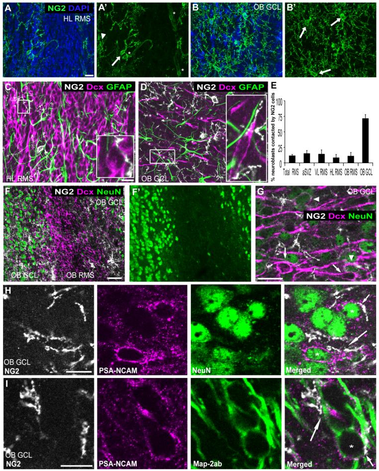
Relation between NG2 cell processes and RMS neuroblasts. A,B: Projected images of z-stacks through 12 μm of P42 mouse sagittal section immunolabeled for NG2 (green) and counterstained with DAPI (blue), showing the density of NG2 cells in the HL RMS (A) and in the OB GCL (B). Arrows indicate NG2 cells in the RMS or the OB GCL. Arrowhead shows a parenchymal NG2 cell at the interface with the RMS. Asterisks show NG2-immunopositive vasculature. The density of NG2 cells is higher in the OB GCL than in the HL RMS. C,D: Projected images of of z-stacks through 1 μm of tissue triple labeled for NG2 (white), DCX (magenta), and GFAP (green). The images were obtained from a portion of the 12-μm z-stack presented in A and B, respectively. Insets in C and D correspond to the boxed areas. Inset in D has been rotated 90° clockwise. Asterisks in D indicate individual DCX-positive neuroblasts. E: Percentages of DCX-positive neuroblasts that were contacted by NG2 cells in different subregions of RMS in P42 mice. F,G: Projected images from a P30 mouse coronal section through the OB triple labeled for NG2 (white), DCX (magenta), and NeuN (green) showing a low-magnification image of the OB RMS and OB GCL (F,F′) and a high-magnification image of the OB GCL (G). Most DCX-positive neuroblasts in the OB RMS lack NeuN expression, whereas, in OB GCL, both NeuN-negative (arrows) and NeuN-positive (arrowheads) neuroblasts are seen. H,I: Relation between maturing PSA-NCAM-immunopositive neurons and NG2 cells in P30 mouse OB GCL. Triple labeling for NG2 (white), PSA-NCAM (magenta), and NeuN (green; H), and for NG2 (white), PSA-NCAM (magenta), and MAP2ab (green; I). NG2 cell processes contact the cell bodies (asterisks and short arrow) as well as neurites (long arrows) of PSA-NCAM and NeuN or PSA-NCAM and MAP2ab double-positive maturing neurons. Scale bars = 20 μm in A,C,G; 50 μm in F; 10 μm in H,I.

