
Figure 1.
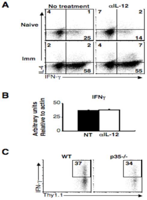
IL-12 at the site of infection is not required for IFN-γ production. (A) Naïve or immune C57BL/6 mice were infected and treated with 1mg αIL-12/23p40 mAb on day -1 and day 3, and on day 5 cells were isolated from infected ears. IL-4 and IFN-γ levels were determined by intracellular cytokine staining. Plots are gated on live CD4+ T cells and are representative of two experiments. Quadrant numbers are shown. (B) RNA was isolated from total ear dermis homogenate, reverse-transcribed and real-time quantitative PCR was performed with primers specific for IFN-γ. Graph shows arbitrary units relative to β-actin. (C) 20 ×106 CD4+ T cells from the spleens and lymph nodes of immune mice were transferred via the retro-orbital plexus into wild-type or IL-12p35-/- mice that were subsequently infected. After 5 days, cells were collected from infected ears and the percentage of IFN-γ+ cells is shown. Plots are gated on live Thy1.1+ donor cells and are representative of two experiments.