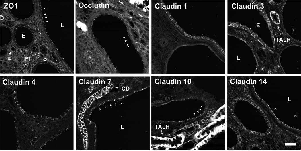
Fig 1.
Expression of tight junction proteins in Pkd1 nl/nl mouse kidney. Immunofluorescence staining was performed on paraffin-embedded sections using antibodies against the indicated proteins, and images acquired by confocal microscopy. Scale bar represents 30 µm. ZO-1: Tight junction staining (arrowheads) of approximately equal intensity is observed in normal, non-cystic tubules such as proximal tubules (PT), and in small (S) and large (L) cysts. Occludin: Occludin is expressed in tight junctions of all cysts, similar to ZO-1. Claudin-1: The upper cyst is a rare cyst staining basolaterally for claudin-1. Most cysts (like the lower cyst in this image) showed no staining at all. Claudin-3: Weak basolateral staining was observed in most cysts, more in small than in large cysts. By comparison, strong claudin-3 staining of a non-cystic thick ascending limb (TALH) is shown. Claudin-4: Basolateral staining was observed in most cysts, more in small than in large cysts. Claudin-7: All cysts showed strong staining for claudin-7, even large cysts such as the one shown here. Note the flattened epithelium and distorted lateral membrane. An intact collecting duct (CD) is also shown. Claudin-10: Occasional small cysts showed weak basolateral claudin-10 staining (arrowheads). By comparison, adjacent non-cystic tubules of the thick ascending limb showed much stronger staining. Claudin-14: Apparent claudin-14 staining of uncertain specificity was observed at the apical membrane (arrowhead) predominantly in very large cysts.

