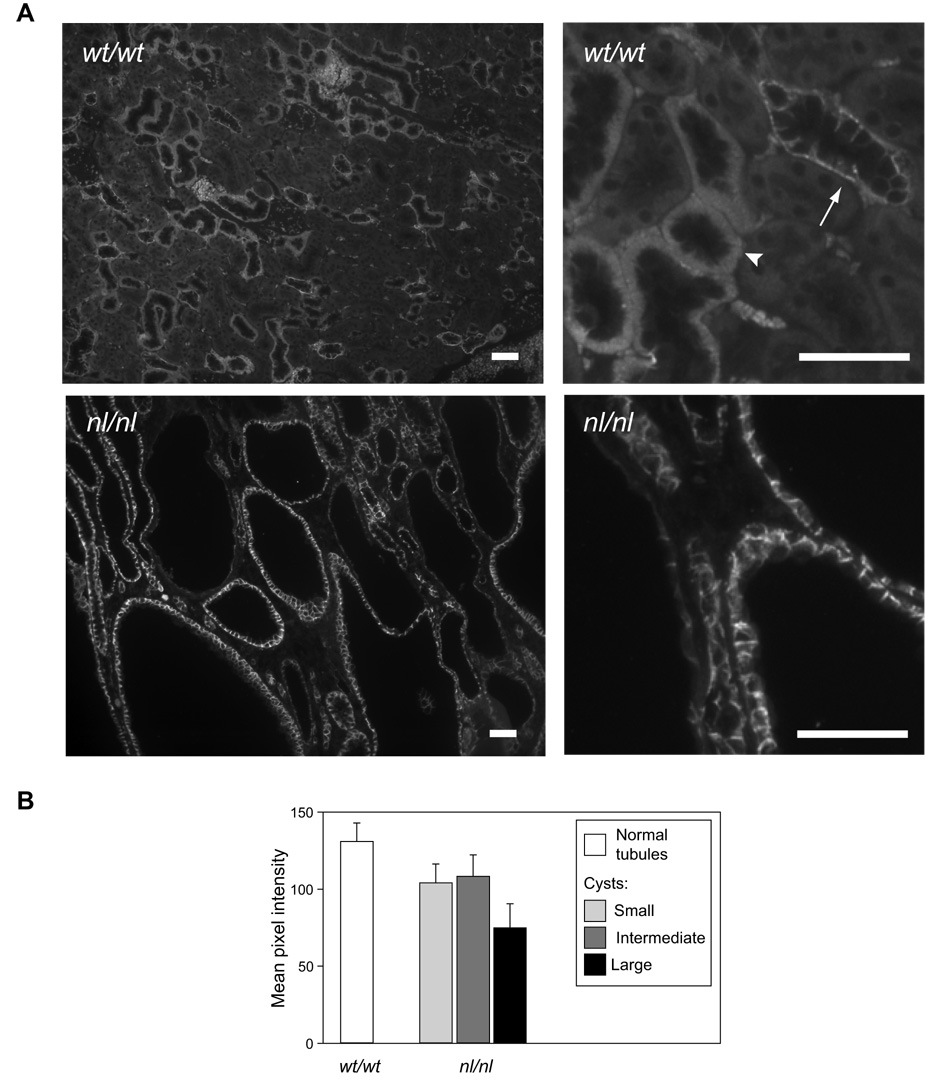
Fig 2.
Relative claudin-7 expression in tubules and cysts. A. The images show low (left) and high (right) magnification views of claudin-7 antibody staining in wild-type (wt/wt), and homozygous (nl/nl) PKD1 hypomorphic mice. Claudin-7 is expressed on the basolateral membrane. In wild-type renal tubules, this appears as a diffuse staining in the thick ascending limb/distal tubule (arrowhead) due to extensive basal membrane invaginations in these segments, and as a more discrete delineation of the base and sides of each cell in the collecting ducts (arrow). The section of cystic kidney overall appears darker but this is simply due to the large area occupied by fluid-filled cyst lumens. The cyst walls, particularly of the small and intermediate cysts, in the nl/nl mice, stain similarly to the walls of normal tubules in the wt/wt mice. Scale bar represents 50 µm. B. Quantitation of claudin-7 immunofluorescence intensity in tight junctions and basolateral membranes of tubule and cyst epithelium. Columns represent mean ± S.E (n = 8 tubules or cysts) and is representative of two such experiments. The p value for comparison between groups is non-significant by one-way ANOVA.

