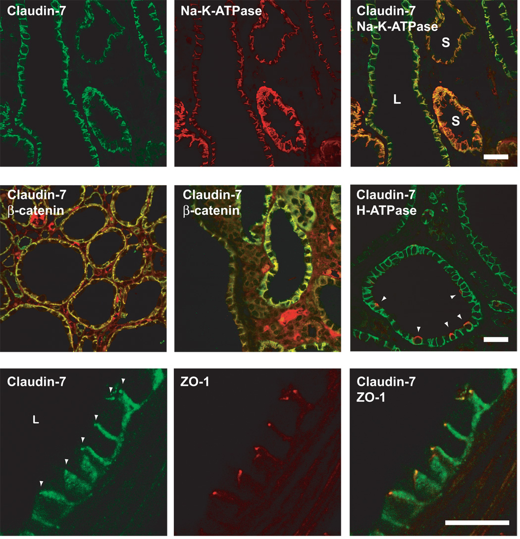
Fig 3.
Double immunofluorescence localization of claudins in Pkd1 nl/nl mouse renal cysts. Scale bars represent 30 µm. Upper row: Double staining for claudin-7 (green) and the α1 subunit of Na-K-ATPase (red) show that both are expressed at the basolateral membrane. Note that the expression of Na-K-ATPase is much lower in large cysts (L) compared to small cysts (S), while claudin-7 expression is well-preserved. This is most obvious in the merged image, where the color of the basolateral membrane in small cysts (orange) clearly differs from that of large cysts (yellow-green). Middle row: Double staining for claudin-7 (green) and β-catenin (red) also shows colocalization to the lateral membrane in cysts of varying size. The apparent interstitial staining is due to non-specific binding of the anti-mouse IgG used as a secondary antibody to detect mouse anti-β-catenin. In the rightmost panel, claudin-7 (green) is demonstrated in a small cyst in which a minority of cells stain at the apical membrane (arrowhead) for H+-ATPase expression (red), representing α-intercalated cells of cortical collecting duct origin. Bottom row: Double staining for claudin-7 (green) and ZO-1 (red) shows that a subset of claudin-7 does localize at the tight junction (arrowheads).

