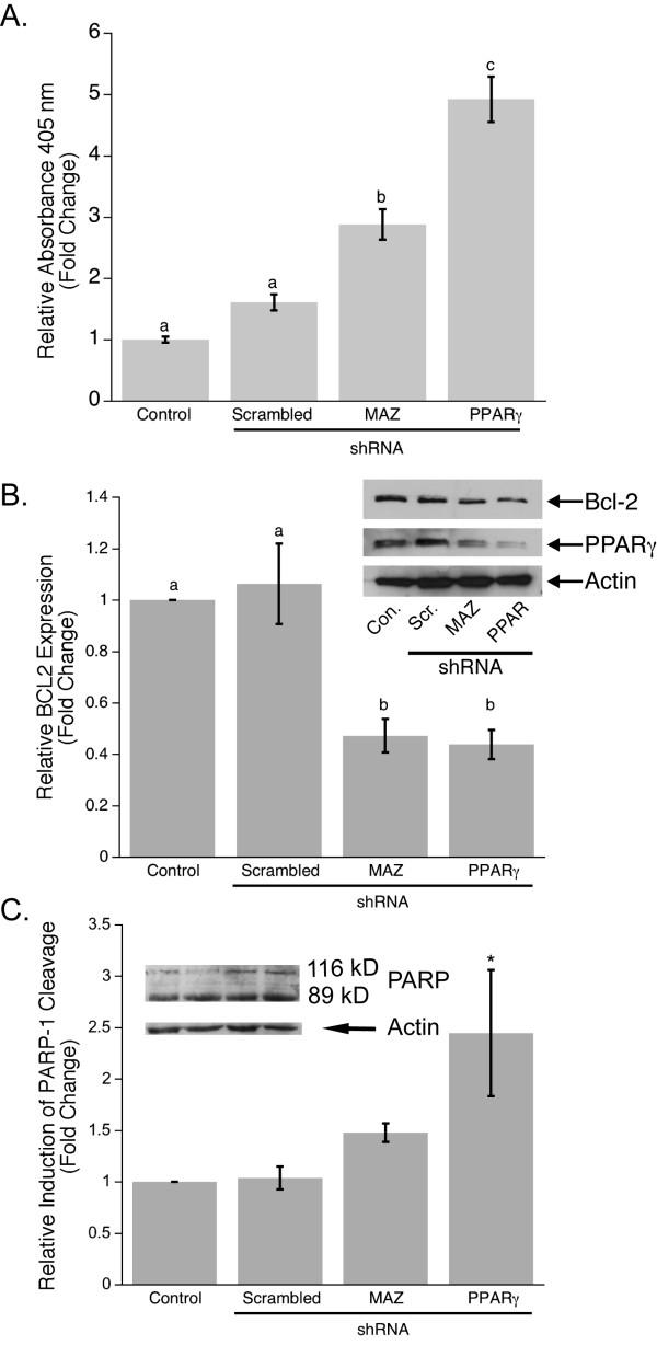
Figure 5.
Down-regulation of PPARγ1 increases apoptosis in MCF-7 breast cancer cells. A. Apoptosis was measured by specific determination of mono- and oligonucleosomes in the cytoplasmic fraction of cell lysates from control and shRNA transfected MCF-7 cells. Data is shown as mean fold changes in cell apoptosis compared to control. Error bars represent s.e.m. and the bars that do not share a letter designation were determined to be significantly different by Tukey's pairwise comparison (p < 0.05). B. Representative Western blot analysis of PPARγ1 and Bcl2 expression in control and shRNA transfected MCF-7 cells showed that a decrease in PPARγ1 expression leads to a decrease in Bcl2 expression. Densitometry was used to quantify Bcl2 expression (n = 4). Bcl2 expression is shown as a fold change in band intensity relative to control MCF-7 cells. Intensity of each band was normalized to actin. C. Western blot analysis of PARP-1 demonstrated an increase in induction of PARP-1 cleavage in MCF-7 cells transfected with PPARγ1 shRNA. Densitometry was used to quantify an 89 kDa fragment of PARP-1 cleavage (n = 3). It is shown as a fold change in the 89 kDa band intensity relative to control MCF-7 cells. Intensity of each band was normalized to actin. * Significantly different from control at p < 0.05.

