Figure 4.
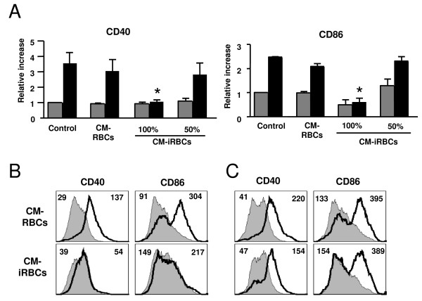
The conditioned medium of P. yoelii-infected erythrocytes inhibits DC maturation. (A) DCs were incubated alone (Control), in the presence of the conditioned media of uninfected (CM-RBCs) or P. yoelii-infected (CM-iRBCs) erythrocytes for 24 h followed by an additional 24 h in the absence (grey bars) or presence (black bars) of 100 ng/ml LPS. The CM-iRBCs was diluted 50% in medium or not (100%). Results are expressed as the relative increase in MFI over DCs incubated in media alone. Error bars represent the standard deviation of 3 averaged independent experiments. *, indicates significant differences in DC surface expression when compared to DC incubated with CM-RBCs and stimulated with LPS. (B) DCs were incubated with either the conditioned medium of uninfected (CM-RBCs) or P. yoelii-infected (CM-iRBCs) erythrocytes in the presence or absence of 100 ng/ml LPS for 24 h. (C) DCs were incubated in the presence of the conditioned medium of uninfected (CM-RBCs) or P. yoelii-infected (CM-iRBCs) erythrocytes for 24 h. DCs were washed and incubated in media alone for 24 h in the presence or absence of 100 ng/ml LPS. (B, C) FACS plots show CD40 or CD86 surface expression on DCs from control cultures (gray filled histogram) or incubated with LPS (black thick line). Mean fluorescent intensity (MFI) values are indicated in each plot for control and LPS stimulated. Each FACS plot is representative of one of at least 2 independent experiments.

