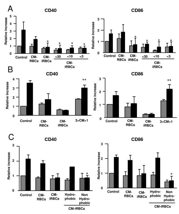
Figure 6.
Characterization of the P. yoelii soluble factor that inhibits DC maturation. (A-C) DCs were incubated alone (Control), in the presence of the conditioned media of uninfected (CM-RBCs) or P. yoelii-infected (CM-iRBCs) erythrocytes for 24 h followed by an additional 24 h in the absence (grey bars) or presence (black bars) of 100 ng/ml LPS. (A) Prior to incubation with DCs, conditioned medium of P. yoelii-infected erythrocytes (CM-iRBCs) was size fractioned using Centricon® filters to smaller than 30 kDa (<30), 10 kDa (<10) and smaller than 3 kDa (<3). (B) Prior to incubation with DCs, molecules of smaller than 1 kDa were dialyzed out of the P. yoelii conditioned medium that was previously size fractioned to smaller than 3 kDa (3>CM>1). (C) Prior to incubation with DCs, The conditioned medium was fractionated based on hydrophobicity using Sep-Pak® columns. (A-C) Results are expressed as the relative increase in MFI over DCs incubated in media alone. Error bars represent the standard deviation of 3 averaged independent experiments. *, represents significant differences in DC surface expression when compared to surface expression on DCs incubated with CM-RBCs and matured with LPS. **, represents significant differences in DC surface expression when compared to surface expression on DCs incubated with CM-iRBCs and matured with LPS.

