
Fig. 6.
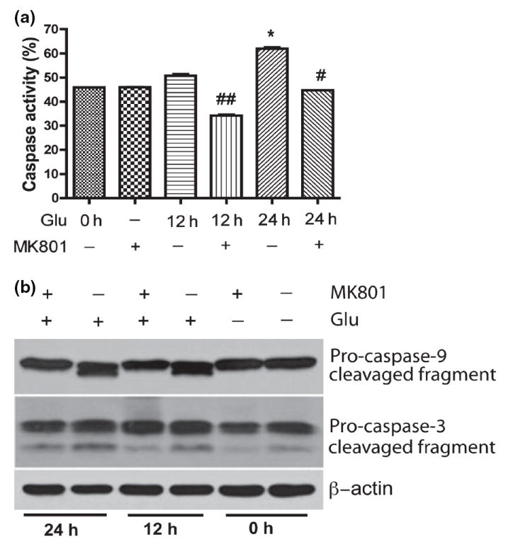
Caspase activation was involved in glutamate-mediated neuronal apoptosis. Rat neurons were treated with 100 μmol/L glutamate and/or 10 lmol/L MK801 as indicated in (a), and then incubated with FITC-VAD-fmk, a FITC-conjugated inhibitory substrate of caspases, and analyzed by Flow Cytometry. The results are expressed as average ±SD of triplicate samples, *denotes p < 0.001 in comparison to control; #denotes p < 0.05 in comparison with glutamate treatment (24 h); ##denotes p < 0.05 in comparison to Glu 12 h. (b) Rat neurons were collected at the indicated times after being treated with 100 μmol/L Glutamate and/or pretreated with 10 μmol/L for 30 min, and were analyzed by western blotting for caspase-9 and caspase-3 cleavage. β-actin was used as a protein loading control.