Figure 5.
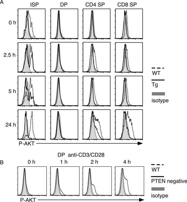
Phosphorylation kinetics of AKT following TCR stimulation. (A) Thymocytes from PTEN transgenic (Tg) or WT littermates were stimulated with plate-bound anti-CD3/CD28 antibodies for the indicated time points. Cells were fixed and stained with antibodies specific for CD4, CD8, TCRβ and phospho-AKT. (B) Thymocytes from PTEN-deficient mice (PTENflox/flox/lck-Cre) [15] or wild-type littermates were stimulated with plate-bound anti-CD3/CD28 antibodies for the indicated time points. Cells were fixed and stained with antibodies specific for CD4, CD8 and phospho-AKT. Cells were analyzed by flow cytometry. The experiments were done more than three times with similar results.

