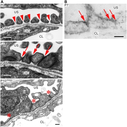
Figure 4. Ultrastructure of mutant podocytes.
(A) Although fine foot processes and slit diaphragms are observed at P0 (arrowheads), the foot processes form irregular adhesions and slit diaphragms are apically mislocalized at P7 (arrows). Mutant podocytes at P10 demonstrates effacement of foot processes and irregular adhesions between foot processes (asteriscs). Apparently, these adhesions did not form tight junctions. The glomerular basement membrane (GBM) is not significantly affected. US, urinary space; EnC, endothelial cell; CL, capillary lumen. Bars, 200 nm. (B) Immunogold electron microscopy in mutant podocytes at P7 reveals that podocalyxin (5-nm gold particles) loses its apically restricted localization, whereas ZO-1 (10-nm gold particles) localizes at the cell–cell junctions of podocytes (arrows). US, urinary space; CL, capillary lumen. Bar, 200 nm.

