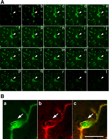
Fig. 5.
Three-dimensional confocal image indicates capillary dysplasia. A: photomicrographs show a series of confocal images of capillary dysplasia morphology at different levels. B: photomicrographs show CD31 and lectin double-labeled immunostaining of dysplasia capillary. Red is CD31-positive endothelial cells, green is lectin-positive capillary walls, and merged yellow color indicates both CD31- and lectin-positive staining. Scale bar = 50 μm.

