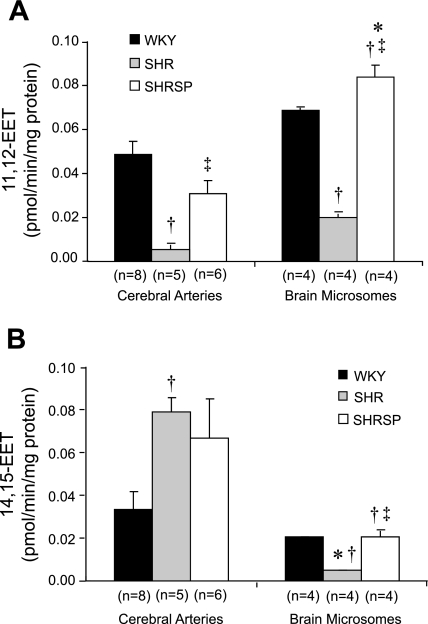
Fig. 4.
Comparison of the formation of 11,12-EET (A) and 14,15-EET (B) in cerebral arteries and microsomes prepared cerebral cortex of SHR, SHRSP, and WKY rats. Mean values ± SE are presented; n, number of animals studied per group. *Significant difference from the corresponding value in cerebral arteries; †significant difference from the corresponding value in WKY rats; ‡significant difference from SHR.

