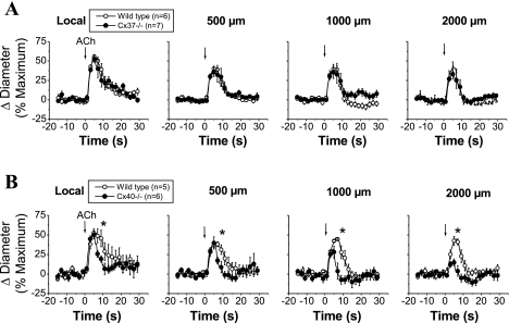
Fig. 3.
Time course of the local and conducted vasodilation induced by ACh in WT, Cx37−/−, and Cx40−/− mice. ACh was ejected by a pressure pulse (300 ms) via a micropipette to stimulate a short segment of the cremasteric arteriole, and the vasodilator response was analyzed at the stimulation site (local) and at locations at 500, 1,000, and 2,000 μm upstream. The vasodilator responses initiated by ACh in two different groups of experiments performed in WT animals were compared with the vasodilation observed in arterioles from Cx37−/− (A) and Cx40−/− (B) animals. Arrows indicate the time at which the stimulus was applied. *P < 0.05 vs. WT mice by one-way ANOVA plus the Newman-Keuls post hoc test.

