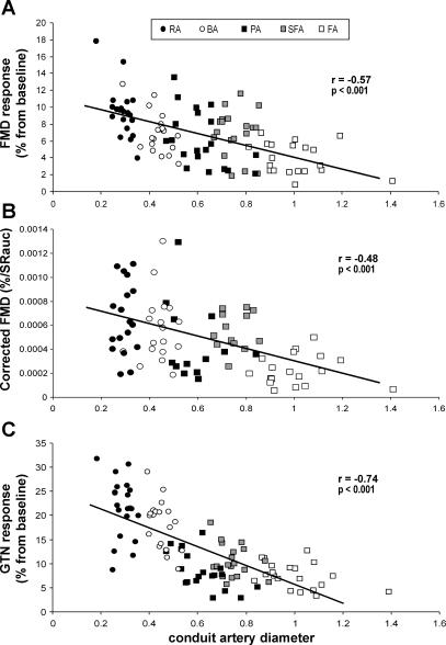
Fig. 1.
Scatter plot for all 5 artery diameters (RA, radial artery; BA, brachial artery; PA, popliteal artery; SFA, superficial femoral artery; FA, common femoral artery) vs. the relative increase from baseline diameter during flow-mediated dilation (FMD; A), the corrected FMD for shear rate area under the curve (SRAUC; B), and the relative change during glyceryl trinitrate (GTN) response (C) in 20 healthy young subjects.

